Consult Corner: Diabetic foot wounds
"Consult Corner" addresses a consult commonly encountered by an on-call resident. The column begins with the reason for consult and assesses questions that might go through a resident's mind as he or she heads to the emergency department to see the patient. Key aspects of the history and physical, as well as additional testing that should be obtained, are also presented. Finally, a review of the decision-making process will present possible management strategies, all of which are synthesized into the context of an actual case.
MESSAGE ON YOUR PAGER:
DIABETIC FOOT WOUND, CALL BACK PLEASE.
You're at an Italian restaurant and about to cut into a serving of steaming lasagna when your pager shows this message from the Medicine service: "Patient has just been admitted with a diabetic foot wound." How should you evaluate and treat this patient?
Vascular
The foot is supplied by the anterior tibial, posterior tibial and peroneal vessels, and normally it has extensive collateral circulation. Each of these vessels gives off smaller perforating arteries on which flaps can be based. The anterior tibial artery continues distally onto the foot as the dorsalis pedis artery lateral to the extensor hallucis longus tendon.
The posterior tibial artery gives rise to cutaneous perforators between the flexor digitorum longus and the soleus as it courses toward the tarsal tunnel. It then gives a medial calcaneal branch before bifurcating into the medial and lateral plantar arteries in the foot. The medial plantar artery then divides into superficial and deep branches – classically, the medial plantar artery flap is based on this superficial branch.
The peroneal artery gives off perforators to supply the posterolateral leg before terminating in an anterior perforating branch and a calcaneal branch.
History
About 25 percent of patients with diabetes get an ulcer. In patients with chronic wounds, it's important to get a sense of the patient's overall medical history and the underlying factors that led to the development of the wound.
Particular aspects of the wound that should be inquired about include: the amount of time the wound has been present; if any preceding, minor trauma was present; prior instances of osteomyelitis or other infections; and prior treatment of the wound. In patients with foot wounds, it's also important to determine if they are ambulatory – and if not, when was the last time that they walked.
General medical issues that should be inquired about include neuropathy (the most important risk factor for development of a diabetic foot wound); diabetic control (hemoglobin A1c); smoking status; any history of vascular disease or related symptoms (claudication, rest pain); and any other major medical comorbidities (e.g., congestive heart failure, chronic obstructive pulmonary disease or chronic kidney disease).
Exam
The lower extremity should be observed for its overall condition (e.g., atrophic, swollen, etc.), and the size and extent of the wound should be noted – including signs of infection and any exposed structures (bone, tendon, etc.). A vascular exam should include palpation of dorsalis pedis and posterior tibial pulses, and if that's abnormal, a pencil Doppler exam should be conducted. Importantly, a "Dopplerable" pulse in one of these vessels may represent retrograde flow through collateral circulation, so consider occluding collateral flow with your finger to ensure you are hearing an antegrade signal in the vessel. A detailed description of this can be found in the references.
A full sensory and motor exam of the foot should be conducted. Bony deformities that include hammer toe and Charcot foot are common in patients with diabetes and should be noted, as these predispose to wounds.
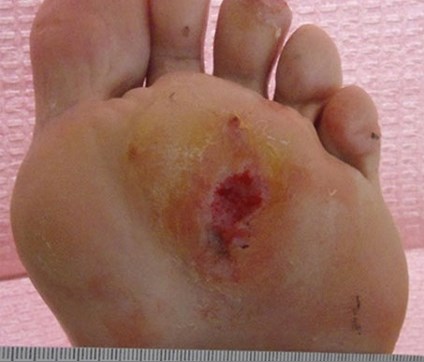
The depth and dimensions of the wound itself should be noted, as well as the presence of any exposed bone and surrounding cellulitis.
Assessment
Diabetic foot wounds often require multidisciplinary management to obtain an optimal outcome. Osteomyelitis is often present, requiring extended antibiotics with Infectious Disease guidance. Peripheral vascular disease is also common in this population; perfusion should be assessed and maximized prior to attempting reconstruction. Diagnostic modalities such as lower-extremity arterial flow studies and CT angiography may be used to evaluate perfusion and reconstructive options. Deformities such as Charcot foot may be managed with casting, or via surgery by foot and ankle surgeons. Optimizing underlying conditions such as these can help maximize the chances of successful soft-tissue reconstruction.
Management
A detailed review of foot-wound reconstruction is outside the scope of this article, but a brief summary of reconstructive options follows:
Split-thickness skin graft
For non-weightbearing portions of the foot
Local flaps
Medial plantar artery flap
Pedicle: superficial branch of medial plantar artery
Notes: This can reach from heel to midfoot. It also has been performed in reverse fashion to reach the forefoot, by raising it on retrograde flow from the dorsalis pedis artery and rotating it 180 degrees.Intrinsic muscle flaps
Small intrinsic muscles of the foot have been used as flaps to cover small defects. These include the abductor digiti minimi, abductor hallucis brevis and extensor digitorum brevis. These can be very useful for small wounds with no other local flap options.
Regional flaps
Reverse sural flap
Pedicle: peroneal artery perforator 5 cm proximal to lateral malleolus and lesser saphenous vein
Notes: This flap is notorious for venous congestion and partial flap loss. It does provide a decent amount of tissue that can be used for wounds around the ankle.
Lateral supramalleolar flap
Pedicle: peroneal artery perforator 5 cm proximal to lateral malleolus Notes: This is a propeller flap based on a distal peroneal artery perforator and is useful for wounds on the lateral ankle and foot.
Propeller flap
Notes: As previously noted, each of the three major vessels to the leg gives off numerous cutaneous perforators as they travel to the foot. Any of these can be used to design a propeller flap. Careful dissection of the perforator is paramount, and it should be ensured that the pedicle does not occlude when the flap is rotated. These can be useful to prevent the need for free tissue transfer.
Free tissue transfer
Many free flaps have been performed on the foot – and this is often the gold standard for distal lower-extremity reconstruction. Individual choices depend on the size, location and other characteristics of the wound in question, as well as the potential donor sites. Especially in patients with vascular disease, it can be prudent to perform the microsurgical anastomosis in end-to-side fashion to preserve as much distal perfusion to the foot as possible.
Conclusion
Foot wounds are very common in patients with diabetes, and as plastic surgeons we are sometimes called to assist in their management. Any assessment of a diabetic foot wound should include an understanding of the "big picture" of the patient's care to ensure that their underlying condition is optimized. The other, arguably more important, aspects of the wound-care regimen – such as proper pressure offloading, treatment of infection and maximization of perfusion – should be optimized in concert with planning for soft-tissue coverage. Despite efforts to resolve diabetic foot wounds, up to 10 percent of patients will ultimately require amputations rather than salvage.
Dr. Aronson is PGY2, and Dr. Kearney is PGY4, in the Northwestern University Feinberg School of Medicine's Division of Plastic Surgery.